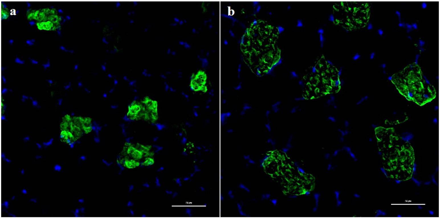
The gastrocnemiusmuscle (GM) of young (3months) and aged (12 months) femalewild-type C57/BL6 micewas examined by light and electron microscopy, looking for the presence of structural changes at early stage of the aging process.Morphometrical parameters including body and gastrocnemius weights, number and type ofmuscle fibers, cross section area (CSA), perimeter, and Feret's diameter of single muscle fiber,were measured.Moreover, lengths of the sarcomere, A-band, I-band, H-zone, and number and CSA of intermyofibrillar mitochondria
IFM were also determined. The results provide evidence that 12 month-old mice had significant changes on skeletal muscle structure, beginning with the reduction of gastrocnemius weight to body weight ratio, compatible with an early loss of skeletal muscle function and strength. Moreover, light microscopy revealed increased muscle fibers size,with a significant increase on their CSA, perimeter, and diameter of both type I and type IImuscle fibers, and a reduction in the percentage of muscle area occupied by type II fibers. Enhanced connective tissue infiltrations, and the presence of centrally nucleated muscle fibers, were also found in aged mice. These changes may underlie an attempt to compensate the loss of muscle mass and muscle fibers number. Furthermore, electron
microscopy discovered a significant age-dependent increase in the length of sarcomeres, I and H bands, and reduction on the overlapped actin/myosin length, supporting contractile force loss with age. Electron microscopy also showed an increased number and CSA of IFMwith age,whichmay revealmore endurance at 12months of age. Together, mice at early stage of aging already showsignificant changes in gastrocnemiusmuscle morphology and ultrastructure that are suggestive of the onset of sarcopenia


