Objective: This study is concerned with clarification of radiological findings that should be addressed and reported in patients listed for cochlear implant (CI) operation. These findings may force a surgeon to consider modifications of the surgical approach by a CI surgeon.
Materials and Methods: The study was performed from January 2015 to January 2016. It included 50 patients with severe‑to‑profound sensorineural hearing loss who fulfilled the criteria for CI. Patients underwent CI surgery in the Department of Otolaryngology. All patients underwent preoperative computed tomography (CT) and magnetic resonance imaging (MRI) assessment in the Department of Diagnostic Radiology. Combined examination of the CT and MRI by the radiologist and the surgeon was advocated.
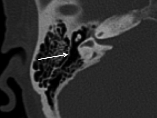
Results: Many anatomical variants were observed regarding the pattern of mastoid pneumatization, position of middle cranial fossa dura, sigmoid sinus position jugular bulb position, and the size and position of the mastoid segment of facial nerve canal. Labyrinthitis ossificans was seen in 3 patients (6%), otospongiosis in 1 patient (2%), and dilated vestibular aqueduct and endolymphatic sac in 9 patients (18%).
Conclusion: Cochlear implantation is a major treatment modality in patients with severe‑to‑profound sensorineural hearing loss. Radiological evaluation is integral in surgery planning.


