

Abstract
Detection of a technique used for diagnosis of Community Acquired Pneumonia by chest
ultrasound compared to chest radiograph.
Study: Sixty two patients presented with clinically diagnosed pneumonia (acute presentation of
fever, cough, purulent expectoration and typical auscultation as rales and bronchial breath sound),
patients with chronic chest and cardiac diseases are excluded from the study. Chest ultrasound and
chest X-ray were done for all patients.
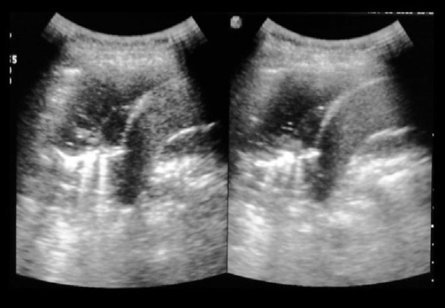
Results: Chest ultrasound showed a significant diagnostic value of consolidation, as it showed
signs of consolidation in 46 patients (74.2%) (P value 0.01), while Chest X-ray showed signs of consolidation
in 32 patients (51.6%) (P value more than 0.05).
Conclusion: Performance of chest ultrasound for the detection of pneumonia is excellent and
superior to chest X-ray considering rapid access to bedside ultrasound and the excellent performance
of this simple test.
Clinical importance: This study supports the routine use of chest ultrasound for the detection of
community acquired pneumonia especially in cases in which chest X-ray is contraindicated or inaccessible.