

Background: Aortic coarctation is a congenital narrowing of the aorta. Computed tomography angiography is used to detect aortic coarctation and determine its extent and severity.
Objective: To assess pediatric aortic coarctation by com-puted tomography (CT) angiography.
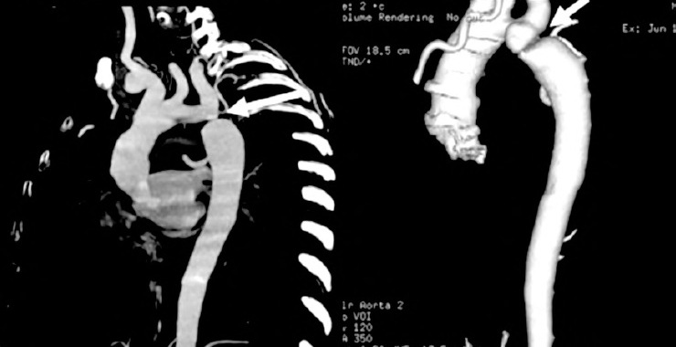
Material and Methods: This study included eighteen pa-tients (10 males and 8 females) who were clinically diagnosed to have coarctation. Both echocardiography and CT angiog-raphy were done to all patients. Axial, multi-planar and volume rendering reconstructions are done to all patients. The presence, site and extent of coarctation are reported. Other cardio-vas-cular anomalies and findings are also registered. The results of both CT angiography and echocardiography are compared to each other.
Results: Seventeen from the examined group were had coarctation of the aorta. The residual patient had double aortic arch. The sensitivity of CT angiography for diagnosis of the coarctation of the aorta was (100%) which was higher than that of Doppler echocardiography (82%). The sensitivity of CT angiography for the assessment of cardiac defects was (72%) which was lower than that of Doppler echocardiography (100%).
Conclusion: CT angiography with reconstruction tech-niques is a valuable imaging modality for pediatric patients with coarctation of the aorta.