

Take home massage
ü Magnetic resonance (MR) imaging is the modality of choice for detecting meniscal injuries and planning subsequent treatment.
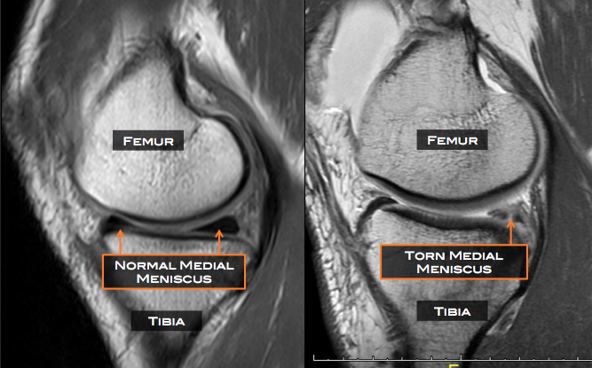
ü Familiarity with the normal anatomy, common anatomic variants , anatomic structures , and indirect secondary signs of meniscal tears can help reduce interpretation errors
üWhen a meniscal tear is identified, accurate description and classification of the tear pattern can guide the referring clinician in patient education and surgical planning. For example, longitudinal tears are often amenable to repair, whereas horizontal and radial tears may require partial meniscectomy.
ü Tear patterns include horizontal, longitudinal, radial, root, complex, displaced, and bucket-handle tears.
üOccasionally, meniscal tears can be difficult to detect at imaging; however, secondary indirect signs, such as a parameniscal cyst, meniscal extrusion, or linear subchondral bone marrow edema, should increase the radiologist’s suspicion for an underlying tear
üMeniscal tears can be treated with conservative therapy, surgical repair, or partial or complete meniscectomy.