

Fawi GH et al.
187
Autonomic and Peripheral Neuropathies in Chronic Liver
Diseases: Clinical and Neurophysiological Study
Fawi GH1
, Khalifa GA1
, Abo Dahab LH2
Departments of Neurology1
, Internal Medicine2
, South Valley University
ABSTRACT
Introduction: Neurological syndromes commonly occur in patients with liver diseases. Neurological deficits
associated with liver diseases may affect the central nervous system, peripheral nervous system, or both. Although
peripheral neuropathy is reported to occur in patients with liver diseases, there is controversy regarding a cause
and effect relationship. Aim of the work: to define the prevalence of peripheral neuropathy and autonomic
neuropathy in patients with chronic liver diseases, to correlate the severity of neuropathy with the severity of liver
diseases, to determine whether neuropathy is related to the cause of liver diseases. Patients and Methods: Sixty
patients with chronic liver diseases, were selected and grouped according to the etiology (viral & non viral) and
severity of liver disease. All patients were subjected to full clinical assessment, liver function tests, blood sugar
and renal function tests, serology using the ELISA technique, abdominal ultrasonography, nerve conduction
studies, electromyography and autonomic function tests. Results: We have found that 45 patients of 60 (75%) had
evidence of neuropathy either clinical and/or electrophysiological, 80% of patients had signs of peripheral
neuropathy, while 45% reported symptoms of peripheral neuropathy, sensory signs and symptoms were reported
more than the motor ones with predilection of the lower limbs, and were related to severity of liver disease.
Autonomic dysfunctions reported in 53% of patients, and the majority of abnormalities were related to patients of
class C. The impairment of nerve conduction reported in high percent of patients of chronic liver diseases
regardless the presence or absence of manifestations and increases with the severity of liver diseases with
predilection of lower limbs. The reported neuropathy was both axonal and demylenating. Conclusion: neuropathy
is very common the nerve conduction study showed that impairment of nerve conduction is present in high percent
of patients of chronic liver diseases regardless the presence or absence of clinical manifestations and increases
with the severity of liver diseases with predilection of lower limbs. Autonomic dysfunctions are common in patients
with chronic liver diseases with predominance of the parasympathetic dysfunctions. Aetiology has little or no
effect on the severity of peripheral neuropathy. (Egypt J. Neurol. Psychiat. Neurosurg., 2005, 42(1): 187-200).
INTRODUCTION
Neurological syndromes commonly occur in
patients with liver diseases. Neurological deficits
associated with liver diseases may affect the
central nervous system, the peripheral nervous
system, or both1
.
The occurrence of a demyelinating peripheral
neuropathy in liver diseases was first described by
Dayan and Williams2
.
Although peripheral neuropathy is reported
to occur in patients with liver diseases, there is
controversy regarding a cause and effect
relationship3
. While some authors doubt the
existence of hepatic neuropathy, others report an
incidence ranging from 19 to 100%. Most authors
agree that the neuropathy seen in liver diseases is
generally mild or sub-clinical but detailed
characterization of the nature of neuropathy are
lacking4
. Additionally, autonomic neuropathy has
been reported in patients with alcoholic and nonalcoholic
liver diseases5,6,7. though this has not
been characterized in the context of peripheral
neuropathy. For many years, previous reports
stated that both liver diseases and neuropathies
Egypt J. Neurol. Psychiat. Neurosurg. Vol. 42 (1) - Jan 2005
188
are systemic disorders in which both liver and
nerves are damaged in the evolution of the
primary process8
. In the last years many reports
suggested that certain neuropathies arise as a
consequence rather than a concomitant of liver
diseases9
.
Aim of the study:
* To define the prevalence of peripheral
neuropathy and autonomic neuropathy in
patients with chronic liver diseases and to
correlate the severity of neuropathy with the
severity of liver diseases.
* To determine whether neuropathy is related
to the cause of liver diseases, and to
determine the relationship between the
prevalence of autonomic neuropathy and
sensory-motor neuropathy in liver diseases.
PATIENTS AND METHODS
Sixty patients with chronic liver diseases,
were evaluated for the presence of peripheral
(sensorimotor) and autonomic neuropathy. Other
causes of peripheral neuropathy as diabetes
mellitus, alcoholism, renal dysfunction and those
with hereditary peripheral neuropathy were
excluded.
The severity of liver diseases was graded
using the Child-Paugh classification (Table 1):
Table 1. Child-Paugh classification10
.
Group designation I II III
1. Hepatic encephalopathy None Mild Severe
2. Ascites None Moderate Severe
3. Serum bilirubin (mg/dl) < 2 2-3 > 3
4. Serum albumin (g/dl) > 3.5 3-3.5 < 3
5. Nutritional state Good Mild malnutrition Severe malnutrition
Parameters under column (I): take score 1,
Parameters under column (II): take score 2,
Parameters under column (III): take score 3.
Patients with score <7 are graded as class A
(mild), Patients with score 7- 9 are graded as class
B (moderate), Patients with score 10-15 are
graded as class C (severe). In our study; 13
patients were class A, 18 patients were class B,
and 29 patients were class C.
All patients were subjected to:
1- Full clinical assessment (general, abdominal
and neurological), the neurological
evaluation including a history and
neurological examination which was relevant
to the peripheral neuropathy. A history to
elicit sensory or motor symptoms of
neuropathy and a neurological examination
testing for pin prick sensibilities, vibration
sense, muscle strength and deep tendon
reflexes.
2- Laboratory evaluation, in the form of:
A- Liver function tests, including liver
enzymes (SGPT & SGOT), total serum
bilirubin and direct bilirubin
B- Blood sugar and renal function tests.
C- Serology using the ELISA technique for
the determination of the cause of the liver
disease (viral and non-viral cause).
3- Abdominal ultrasonography was done for all
patients to assess the liver size, echogenicity,
portal vein diameter, splenic size and
presence of ascites.
4- Nerve conduction studies were done for the
right and left median nerves, right and left
ulnar nerves and right and left common
peroneal nerves.
Fawi GH et al.
189
- For sensory nerves (median and ulnar)
distal latencies, sensory conduction
velocities and peak to peak amplitude
were measured.
- For motor nerves (median, ulnar and
common peroneal) distal latencies, motor
conduction velocities, baseline to peak
amplitude and F-wave latencies were
measured.
5- E.M.G- was done in abductor policis muscle
bilaterally during rest, minimal contraction,
maximum contraction to detect denervation
pattern as positive sharp wave, fibs, giant
waves and interference pattern.
6- Autonomic function tests included
determination of blood pressure in response
to standing, heart rate variations to rapid
deep respirations and Valsalva maneuver.
The presence of neuropathy was defined by
the presence of abnormalities in at least 2 of 4
categories of symptoms, signs, NCS and
autonomic function tests.
T-test and Chi square test were used for
comparing our groups of data, P-value less than
0.05 is considered as significant and less than 0.01
is considered as highly significant.
RESULTS
Table 2. Demographic data.
Class A Class B Class C
P-value between
A & B A & C B & C
Number of patients (%) 13 (21.67%) 18 (30%) 29 (48.33%)
Age (Mean+SD) 44.2±13.1 50.5±12.8 48.4±9.6 0.19 0.24 0.53
Sex %
Male 61.5% 55.5% 58.6% 0.3 0.6 0.2
Female 38.4% 44.4% 41.3%
Occupation
Manual Worker 30.7% 33.3% 41.3% 0.7 0.1 0.2
Employee 15.3% 5.5% 3.4% 0.03 0.003 0.3
Farmer 23.07% 16.6% 13.79% 0.2 0.1 0.5
Housewife 30.7% 44.4% 41.3% 0.05 0.14 0.98
Duration of illness
in months (Mean±SD
17.07±15.4 32±36.6 23.4±18.9 0.13 0.29 0.29
Number of patients
with PN. (%)
3
(23.07%)
15
(83.3%)
27
(93.1%) 0.003 0.000 0.03
Egypt J. Neurol. Psychiat. Neurosurg. Vol. 42 (1) - Jan 2005
190
Table 3. Classification of patients according to etiology of liver disease.
Non viral HCV HCV&HBV HBV
Number of patients (%)
13
(21.6%)
38
(63.3%)
5
(8.3%)
4
(6.6%)
Table 4. Clinical and Autonomic Manifestations in Different. Classes.
Class
A
Class
B
Class
C
p-value between
A & B A & C B & C
Symptoms
Tingling and numbness in UL 15.3% 33.3% 49.1% 0.003 0.000 0.02
Tingling and numbness in LL 15.3% 38.8% 53.5% 0.002 0.000 0.03
Cramps in UL 0 33.3% 50.9% 0.000 0.000 0.01
Cramps in LL 0 38.8% 49.9% 0.000 0.000 0.08
Weakness in UL 0 5.5% 10.3% 0.01 0.001 0.3
Weakness in LL 0 5.5% 10.3% 0.01 0.001 0.3
Signs
Glove hypothesia 30% 38.8% 51.7% 0.18 0.001 0.06
Stock hypothesia 30% 61% 74.2% 0.0001 0.000 0.05
Diminished or lost deep reflexes
Upper limbs 30% 44.4% 59.5% 0.04 0.0002 0.02
Knee 46.1% 61% 75.8% 0.03 0.0000 0.02
Ankle 53.8% 77.7% 82.7% 0.0003 0.0000 0.4
Deep sensory loss UL 15.3% 16.6% 24.1% 0.8 0.07 0.14
Deep sensory loss LL 23% 23.9% 24.1% - 0.9 0.9
Distal weakness UL 0 0 6.9% - 0.007 0.007
Distal weakness LL 0 0 6.9% - 0.007 0.007
Muscle wasting 0 0 0 - - -
Autonomic dysfunction 15.3% 72.2% 76.4% 0.0000 0.000 0.5
Fawi GH et al.
191
Table 5. Motor Conduction Study.
Rt. Side Lt. Side
DL MCV Amp. F-wave DL MCV Amp. F-wave
26
(43.3%)
21
(35%)
20
(33.3%)
19
(31.6%)
28
(46.6%)
17
(28.3)
20
(33.3%)
19
(31.6%)
Median n.
4
(30.7%)
2
(15.3%)
5
(38.4%)
2
(15.3%)
6
(46.1%)
0
(0%)
4
(30.7%)
4
(30.7%)
Class A
7
(38.8%)
9
(50%)
6
(33.3%)
7
(38.8%)
7
(38.8%)
4
(22.2%)
6
(33.3%)
4
(22.2%)
Class B
15
(51.7%)
10
(34.5%)
9
(31.1%)
10
(34.5%)
15
(51.7%)
13
(44.8%)
10
(34.5%)
11
(37.9%)
Class C
Pv (A & B) 0.2 0.7 0.000 0.09 0.002 0.8 0.000 0.09
Pv (A & C) 0.18 0.6 0.000 0.08 0.003 0.18 0.003 0.02
Pv (B & C) 0.041 0.9 0.003 0.05 0.8 0.96 0.02 0.05
15
(25%)
10
(16.6%)
21
(35%)
8
(13.3%)
19
(31.6%)
8
(13.3%)
29
(48.3%)
1
(1.6%)
Ulnar n.
1
(7.6%)
1
(7.6%
3
(23%)
0
(0%)
4
(30.7%)
0
(0%)
4
(30.7%)
0
(0%)
Class A
4
(22.2%)
3
(16.6%)
5
(27.7%)
1
(5.5%)
4
(22.2%)
2
(11.1%)
8
(44.4%)
0
(0%)
Class B
10
(34.4%)
6
(20.7%)
13
(44.8%)
7
(24.1%)
11
(37.9%)
6
(20.7%)
17
(58.6%
1
(3.44%)
Class C
Pv (A & B) - 0.04 0.001 0.06 0.01 0.6 0.05 0.02
Pv (A & C) 0.003 0.0003 0.000 0.07 0.000 0.003 0.02 0.06
Pv (B & C) 0.003 0.02 0.02 0.02 0.006 0.03 0.4 0.01
33
(55%)
49
(81.6%)
51
(85%)
16
(26.6%)
33
(55%)
43
(71.6%)
50
(83.3%)
18
(30%)
Common P. n.
5
(38.4%)
8
(61.5%))
11
(84.6%)
2
(15.3%)
6
(46.1%)
5
(38.4%)
8
(61.5%)
3
(23%)
Class A
9
(50%)
16
(88.8%)
16
(88.8%)
6
(33.3%)
8
(44.4%)
16
(88.8%)
14
(77.7%)
8
(44.4%)
Class B
19
(65.5%)
25
(86.2%)
24
(82.75%)
8
(27.6%)
17
(58.6%)
22
(75.8%)
28
(96.5%)
7
(24.1%)
Class C
Pv (A & B) 0.003 0.04 0.000 0.4 0.003 0.4 0.03 0.006
Pv (A & C) 0.9 0.009 0.001 0.05 0.008 0.8 0.04 0.003
Pv (B & C) 0.003 0.01 0.09 0.04 0.6 0.1 0.5 0.81
Egypt J. Neurol. Psychiat. Neurosurg. Vol. 42 (1) - Jan 2005
192
Table 6. Sensory Conduction Study.
Rt. Side Lt. Side
DL SCV Amp. DL SCV Amp.
Median n.
22
(36.6%)
51
(85%)
7
(11.6%)
18
(30%)
47
(78.3%)
5
(8.3%)
Class A
2
(15.3%)
10
(76.9%)
2
(15.3%)
2
(15.3%)
9
(69.2%)
1
(7.6%)
Class B
6
(33.3%)
15
(83.3%)
1
(5.5%)
6
(33.3%)
13
(72.2%)
0
(0%)
Class C
14
(48.3%)
26
(89.6%)
4
(13.7%)
10
(34.5%)
25
(86.2%)
4
(13.7%)
Pv (A & B) 0.003 0.3 0.03 0.003 0.5 0.007
Pv (A & C) 0.000 0.2 0.4 0.003 0.2 0.07
Pv (B & C) 0.02 0.1 0.05 0.9 0.3 0.000
Ulnar n.
22
(36.6%)
21
(35%)
2
(3.3%)
21
(35%)
26
(43.3%)
3
(5%)
Class A
2
(15.3%)
2
(15.3%)
0
(0%)
3
(23%)
3
(23%)
0
(0%)
Class B
7
(38.8%)
5
(27.7%)
1
(5.5%)
8
(44.4%)
3
(16.6%)
3
(16.6%)
Class C
13
(44.8%)
14
(48.3%)
1
(3.44%)
10
(34.5%)
20
(68.9%)
0
(0%)
Pv (A & B) 0.002 0.05 0.01 0.003 0.14 0.000
Pv (A & C) 0.000 0.000 0.003 0.04 0.0001 -
Pv (B & C) 0.4 0.007 0.3 0.2 0.000 0.000
Fawi GH et al.
193
Table 7. Clinical and autonomic manifestations in non viral and post HCV chronic liver diseases.
Non viral HCV p-value between non
viral & post. HCV
Symptoms
Tingling and numbness in UL 23.1% 30.6% 0.2
Tingling and numbness in LL 23.1% 26.2% 0.6
Cramps in UL 11.6% 16.2% 0.3
Cramps in LL 11.6% 15.9% 0.3
Weakness in UL 7.7% 5.2% 0.3
Weakness in LL 7.7% 5.2% 0.3
Signs
Glove hypothesia 46.1% 39.4% 0.3
Stock hypothesia 46.1% 55.2% 0.2
Diminished or lost deep reflexes
Biceps 53.8% 39.4% 0.03
Brachioradialis 53.8% 39.4% 0.03
Triceps 53.8% 39.4% 0.03
Knee 76.9% 52.6% 0.003
Ankle 76.9% 63.1% 0.3
Deep sensory loss UL 23% 10.5% 0.02
Deep sensory loss LL 30.7% 10.5% 0.001
Distal weakness UL 7.6% 5.2% 0.3
Distal weakness LL 7.6% 5.2% 0.3
Muscle wasting 0 0 -
Autonomic dysfunction 38.4% 44.3% 0.4
DISCUSSION
In our study we prospectively evaluated 60
patients with chronic liver diseases to assess the
presence of peripheral and autonomic
neuropathies regarding their types, prevalence,
relation to severity and etiology of liver diseases.
13 patients were in Class A, 18 patients were in
class B and 29 patients were class C, as shown in
table (3) .Patients with liver diseases of non viral
causes were 13(21.6%) and those due to viral
causes (virus C, virus A, virus B) were 47patients
(78.4%) .Manual workers were more reported in
class C 41.3%,while employee were more in class
A. 15.35%.
Most authors agree that the neuropathy seen
in liver disease is generally mild or subclinical but
detailed characterization of the nature of
neuropathy are lacking4
.Additionally, autonomic
neuropathy has been reported in patients with
alcoholic and non-alcoholic liver disease5,6,7
though this has not been characterized in the
context of peripheral neuropathy.
There has been no systematic
characterization of peripheral neuropathy in the
context of coexisting liver failure4
. Regarding
neurological findings, we reported that 48 patients
(80%) were presented with signs of peripheral
neuropathy in the form of glove hypoesethia,
stocke hypoesethia, diminished or lost deep
reflexes, deep sensory loss, and lastly distal
Egypt J. Neurol. Psychiat. Neurosurg. Vol. 42 (1) - Jan 2005
194
weakness. Our findings regarding signs of
peripheral neuropathy were reported more in
lower limbs than upper limbs, more in patients
with viral causes, and were related to severity of
liver diseases, where in class C reported in 93% of
patients, in 83% in class B, and in only 23% of
patients in class A.
As shown in table (4), glove hypoesthesia
was present in 30% of patients of class A, 38.8%
of patients of class B and in 51.7% of patients of
class C, As regard stock hypoesthesia, it was
present in 30% of patients of class A, 61% of
patients of class B and in 74.2% of patients of
class C, Lost or diminished deep reflexes were
detected in the upper limb in 30% of patients of
class A, in 44.4% of patients of class B and in
59.5% of patients of class C, lost or diminished
knee reflexes were detected in 46.1% of patients
of class A, in 61% of patients of class B and in
75.8% of patients of class C, lost or diminished
ankle reflexes were detected in 53.8% of patients
of class A, in 77.7% of patients of class B and in
82.7% of patients of class C, deep sensory loss in
the upper limbs was detected in 15.3% of patients
of class A, in 16.6% of patients of class B, and in
24.1% of patients of class C, as regard deep
sensory loss in the lower limbs, it was detected in
23% of patients of class A, in 23.9% of patients of
class B, and in 24.1% of patients of class C, distal
weakness either in the upper and lower limbs was
detected in only 6.9% of patients of class C. No
muscle wasting was detected in patients of the
study.
From the above results we conclude that
signs of peripheral neuropathy are more frequent
in patients with more severe liver disease, also the
lower limbs are affected more than the upper
limbs.
These results are in agreement but higher
than a previous study11 which reported that at
neurological examination of cirrhotic patients a
sensitive distal deficit was clinically observed in
25.8% of patients, a distal motor involvement in
41.9%, and a mixed sensorimotor impairment in
6.5%. Deficit was mainly appreciable in the lower
limbs.
Our results are also in agreement but higher
than another study12 which reported that clinical
signs of peripheral nerve involvement were found
in 21% of patients with chronic liver diseases.
Another study4
reported that 29% of patients had
abnormalities on examination consisting of distal
sensory loss to vibration in 20.6% patients, and
pin in 12.6% of patients; reduced or absent ankle
reflexes in 20.6%of patients; and toe extensor
weakness in 1.7% of patients. In accordance with
our results one of the Egyptian studies at 200213
,
signs of peripheral neuropathy were reported in
86.7% of there patients that agree with our results.
Regarding symptoms of peripheral
neuropathy (table 4), 45% of patients were
presented with mild non disabling symptoms in
the form of mild cramps, paraesethia, and
numbness, and only 6.6% of patients were
presented with distal weakness. The sensory
symptoms were more common than the motor
ones. These symptoms were reported in lower
limbs more than upper limbs, and were related to
severity of liver disease, where in class C 49.1%
of patients were suffering from the previous
symptoms compared with 15.3% of patients in
class A. Distal paresthesias (tingling and
numbness) in upper limbs were present in 15.3%
of patients of class A, in 33.3% of patients of
class B and in 49.1% of patients of class C,
whereas distal paresthesias in lower limbs were
present in 15.3% of patients of class A, in
38.8%of patients of class B and in 53.5% of
patients of class C. Distal cramps in upper limbs
were not present in patients of class A, present in
33.3% of patients of class B and in 50.9% of
patients of class C, also distal cramps in lower
limbs were not present in patients of class A,
present in 38.8% of patients of class B and in
49.9% of patients of class C. None of the patients
related to class A reported history of distal
weakness neither in the upper limbs nor in the
lower limbs, however, 5.5% of patients in class B
had reported distal weakness in both upper and
lower limbs. As regard class C 10.3% of the
patients had reported distal weakness in both
upper and lower limbs. These results show
Fawi GH et al.
195
obvious increase in the symptoms with the
increase of the severity of the liver disease with a
highly statistically significant difference in the
majority of them. Our results are in agreement
but higher than that of a previous study.4
, which
reported that 22% of patients had symptoms of
neuropathy consisting of mild cramps,
paraesthesia, or numbness. Also our results are
higher than another one13 which reported that
26.7% of their patients were suffering from
symptoms of peripheral neuropathy.
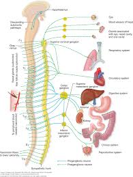
Regarding autonomic neuropathy, autonomic
dysfunctions were reported in 53.3% of our
patients in varying degrees according to severity
of liver disease. Several autonomic function tests
were done for all patients and showed that one or
more of all tests used was found to be abnormal in
15.3% of patients of class A, in 72.2% of patients
of class B and in 76.4% of patients of class C.
Abnormal heart rate variations in response to
rapid deep respiration were found in 23% of
patients of Class A, in 61.1% of patients of Class
B and in 72.4% of patients of Class C. As regard
abnormality in heart rate variations in response to
Valsalva maneuver it was found in 15.3% of
patients of Class A, in 44.4% of patients of Class
B and in 68.9% of patients of Class C. Abnormal
blood pressure response to standing was present in
7.6% of patients of Class A, in 16.6% of patients
of Class B and in 17.2% of patients of Class C.
No patient with abnormal autonomic function tests
(AFTs) had symptoms related to autonomic
dysfunction. Autonomic dysfunctions were
reported in patients with viral liver diseases
61.6% more than those of non viral causes 38.4%.
From our results we found that autonomic
dysfunctions are common in patients with chronic
liver diseases and the majority of abnormalities
were related to patients of class C, followed by
those of class B and lastly those of class A, and
the differences were statistically significant.
Our result agree with those of Bajaj et al.14
,
who reported that autonomic dysfunction was
significantly more frequent in advanced liver
disease compared with early liver damage. Nine
(75%) out of 12 cirrhotic subjects belonging to
Child class B and six (85.7%) of the seven
patients belonging to Child class C had autonomic
neuropathy while only one patient with Child class
A had early parasympathetic damage, and also
with those of McDougall et al.15
, who reported
that autonomic neuropathy were common
(occurring in 50%) in patients with endstage liver
disease and were more frequent than previously
reported. One of the previous studies16
. reported
that autonomic neuropathy is common in alcoholic
patients but the fact that it is found with
comparable frequency in non-alcoholic liver
disease suggest that the neurological defect may
be secondary to the disturbed liver function.
Also in agreement with our results
Hendrikse et al.17
, reported that, autonomic
impairment was closely associated with hepatic
function with 23% of Child-pugh class A and 69%
of class B and C. Also shaza et al.
13
, reported
63.3% of their patients were suffering from
autonomic dysfunctions. Returning to our results,
we can notice that, most of the autonomic
dysfunctions reported were in the form heart rate
variations denoting predominant parasympathetic
dysfunctions in patients with chronic liver
diseases, this notice are supported by previous
results5,13
.
Nerve Conduction Study (tables 5 & 6):
Patients with abnormal sensory conduction
study(median nerve) were 85% of all patients
compared with 33.3% with abnormal motor
conduction study with significant statistical
difference, while the motor conduction study of
the common peroneal nerve was more affected
(83.3%90) than that of nerves of the upper limbs.
(a) Distal latency: prolonged motor and sensory
distal latencies were very common in the
median nerve and entrapment neuropathy
were detected in about 35.7% of patients,
these findings agree the results of Chaudhry
et al.4
, who reported that entrapment
neuropathy can occur in about one third of
patients.
Prolonged motor distal latency of the
ulnar nerve was a rare finding, occurring in
Egypt J. Neurol. Psychiat. Neurosurg. Vol. 42 (1) - Jan 2005
196
the right ulnar nerve in 1.6% of patients of
and in the left ulnar nerve in 13.3% of
patients. On the other hand the prolonged
sensory distal latency of the ulnar nerve was
found in 36.6% of patients on the right side
and in 35% of patients on the left side.
Prolonged motor distal latency of the right
common peroneal nerve were found in 30%
of patients and in 26.6% on the left side. The
incidence of prolonged distal latency was
found to be increased with the severity of
liver disease with the highest incidence in
class C patients and the least incidence in
class A patients. And the difference was
statistically significant.
(b) Conduction Velocity: the highest incidence
of reduced motor conduction velocity(MCV)
was found in both right (83.3% of patients)
and left (85% of patients) common peroneal
nerves, and this agree the results of Lani et
al.11
, who reported that, in
neurophysiologically evaluated patients with
chronic liver disease, at least 87% of patients
featured different degrees of nerve
impairment, mostly located in peroneal
nerves.
Reduced MCV was found in 33.3% of
patients in the right median nerve, in 33.3%
in the left median nerve, 48.3% of patients in
the right ulnar nerve, and in 35% of patients
in the left ulnar nerve. However, reduced
sensory conduction velocity (SCV) was
found in 85% of patients in the right median
nerve, 78.3% of patients in the left median
nerve, 35% of patients in the right ulnar
nerve and in 43.3% of patients in the left
ulnar nerve. Also the incidence of reduced
conduction velocity was found to be
increased with the severity of liver disease
with the highest incidence in class C patients
and the least incidence in class A patients.
And the difference was statistically
significant.
(c) Amplitude: the highest incidence of reduced
motor amplitude was found in both the right
(71.6% ) and left (81.6% ) common peroneal
nerves. However it was reduced in 28.3% of
patients in the right median nerve, in 35% of
patients in the left median nerve,13.3% of
patients in the right ulnar nerve and in 16.6%
of patients in the left ulnar nerve.
Sensory amplitude was reduced in
11.6% of patients in the right median nerve,
in 8.3% of patients in the left median nerve,
in 3.3% of patients in the right ulnar and in
5% of patients in the left median nerve.
Again, the incidence of reduced amplitude
was found to be increased with the severity
of liver disease with the highest incidence in
class C patients and the least incidence in
class A patients. And the difference was
statistically significant.
(d) F-wave latency: the highest incidence of
prolonged F-wave latency was detected in
common peroneal nerves of both sides (55%)
,followed by the right (46.6%) and left
(43.3%) median nerves and lastly the right
(31.6%) and left (25%) ulnar nerves,
however the incidence was more with the
severe cases of liver diseases, but the
difference was statistically significant in few
cases. The presence of prolonged F-wave
latency denote the presence of demyelinating
neuropathy.
Our results are in agreement with those of
Kharbanda et al.18
, who reported that nerve
conduction studies were abnormal in 73% patients
with liver cirrhosis, and with those of McDougall
et al.15
, who reported that nerve conduction study
was abnormal in 93% of patients with chronic
liver diseases and increase with the severity of
liver diseases, also agree those of Fierro et al.
12
,
who reported that clinical signs of peripheral
nerve involvement were found in 21% of patients
with chronic liver diseases while
electrophysiological impairment were found in
57.8% of patients.
These results are also in agreement with
Chaudhry et al.4
, who reported that in patients
with chronic liver diseases 43% had reduced
sensory or motor amplitude on the NCS. The most
Fawi GH et al.
197
common abnormality was reduced or absent sural
sensory amplitudes (in 34%), followed by reduced
peroneal amplitudes (in 26%). Median sensory
amplitude was reduced in 16 % motor amplitude
was decreased in 3%. Peroneal conduction
velocity was reduced in 24% of patients. In
accordance with our results also, Shaza et al.
13
,
reported 60.5% of their patients with sensory
conduction abnormality, and 41.7% with motor
conduction abnormality.
Electromyographic Findings:
Table 8. Percentage of patients with Naturopathic EMG changes in Different Classes.
Class A Class B Class C P-value between
A & B A & C B & C
Neuropathic
EMG changes 46.1% 61.1% 68.9% 0.03 0.02 0.05
Neuropathic electromyographic (EMG)
changes in the form of fibrillations and positive
waves at rest, giant wave on moderate contraction
and poor interference was detected in many cases
of our study. In class A 46.1% of patients had
neuropathic EMG all of them had giant waves on
minimal contraction, 30.1% had poor interference
pattern but none had abnormal findings at rest. As
regard class B, 61.1% of patients had neuropathic
EMG changes 11.1% of patients had fibrillations
at rest, 50% of patients had giant waves on
minimal contraction and 55.5% of patients had
poor interference pattern. 68.9% of patients of
class C had neuropathic EMG changes, 20.6% of
patients had fibrillations at rest, 62% of patients
had giant waves on minimal contraction and
65.5% of patients had poor interference pattern.
The differences between class A & B and class A
& C were statistically significant (p=0.03 and
p=0.02 respectively). but the difference between
class B & C was statistically insignificant
(p=0.05).
The presence of neuropathic EMG changes
increases with the increased severity of liver
disease with statistically significant difference.
The pathogenesis of peripheral neuropathy with
liver disease was discussed in the last few years.
Some systemic disorders that cause liver disease
also are independent causes of peripheral nerve
dysfunction, e.g. alcohol induced cirrhosis,
porphyria, polyarteritis nodosa, amylodosis, and
certain intoxications, all cause independent liver
disease and peripheral neuropathy3,19, so a cause–
effect relationship between both conditions had
been questioned. In our study all of these
conditions had been excluded.
The presence of neuropathic EMG reflects
the axonal element in the pathogenesis of
neuropathy (reported in 46.1% up to 68.9% of
patients), while the prolongation of F-wave
latencies (reported in 25% up to 55% of patients)
reflects the demyelinating pattern of neuropathy,
so the neuropathy of chronic liver disease is both
axonal and demyelinating, however the axonal
element is the predominant.
Our results agree in agreement with those of
Kharbanda et al.18
, who reported that nerve
conduction studies were abnormal in 73% patients
with liver cirrhosis and the pattern of involvement
was predominantly of an axonal sensory motor
polyneuropathy.
Also our results are in agreement with those
of Ripault et al.20
, who reported that the
prevalence of peripheral neuropathy in chronic
liver diseases is 8% and it is an axonal neuropathy
with diminution of the myelinated fibers. And also
are in agreement with those of Lani et al.
11, who
reported that the neurological impairment in
patients with chronic liver diseases is due to
demyelinating inflammation which led to
segmental demyelination and sever loss large
myelinated fibers.
Egypt J. Neurol. Psychiat. Neurosurg. Vol. 42 (1) - Jan 2005
198
Relation of the neuropathy to the etiology of
liver disease
In comparing the incidence of peripheral
neuropathy in non-viral and post HCV cirrhosis,
we have found that:
(A) Symptoms. Distal paresthesia of upper limbs
was present in 23.1% of patients with non
viral chronic liver disease and in 30.6% of
patients with post HCV cirrhosis. The
percentage of the presence of distal
paresthesia of lower limbs in patients with
non viral chronic liver disease was (23.1%)
and of patients with post HCV cirrhosis was
(26.2%). 11.6% of patients with non viral
aetiology were presented with cramps in the
upper limbs in comparison with 16.2% of
patients with post HCV cirrhosis. As regard
the cramps in the lower limbs they were
present in 11.6% of patients with non viral
aetiology and in 15.9% of patients with post
HCV cirrhosis. 7.7% of patients with non
viral aetiology have reported history of distal
weakness in the upper limbs and 5.2% of
patients with post HCV cirrhosis. Also 7.7%
of patients with non viral aetiology have
reported history of distal weakness in the
lower limbs in comparison with 5.2% of
patients with post HCV cirrhosis.
(B) Signs: Glove hypoesthesia was present in
46.1% of patients with non viral chronic liver
disease, and in 39.4% of patients with post
HCV cirrhosis. As regard stock hypoesthesia,
it was present in 46.1% of patients with non
viral chronic liver disease and in 55.2% of
patients with post HCV cirrhosis. Lost or
diminished deep reflexes were detected in the
upper limb (biceps, brachioradialis and
triceps) in 53.8% of patients with non viral
chronic liver disease and in 39.4% of patients
with post HCV cirrhosis and. Lost or
diminished knee reflexes were detected in
76.9% of patients with non viral chronic liver
disease and in 52.6% of patients with post
HCV cirrhosis. Lost or diminished ankle
reflexes were detected in 76.9% of patients
with non viral chronic liver disease and in
63.1% of patients with post HCV cirrhosis.
Deep sensory loss in the upper limbs was
detected in 23% of patients with non viral
chronic liver disease and in 10.5% of patients
with post HCV cirrhosis. As regard deep
sensory loss in the lower limbs was detected
in 24.1% of patients with non viral chronic
liver disease and in 10.5% of patients with
post HCV cirrhosis. Distal weakness either in
the upper and lower limbs was detected 7.6%
of patients with non viral chronic liver
disease and in 5.2% of patients with post
HCV cirrhosis .From the above results we
have found that no significant differences as
regard the presence of symptoms and signs of
peripheral neuropathy between patients with
non viral chronic liver disease and patients
with post HCV cirrhosis.
(C) NCS & EMG: The same as clinical
manifestations NCS &EMG in patients with
non viral chronic liver disease and patients
with post HCV cirrhosis has no significant
difference. And so no relation has been found
between the aetiology of liver disease and the
presence of neuropathy.
(D) Autonomic dysfunction: Autonomic
dysfunction was fount in 38.4% of patients
with non viral chronic liver disease and in
44.3% of patients with post HCV cirrhosis
with no statistically significant difference
between the two groups. So, the presence of
autonomic dysfunction has no relation to the
aetiology of liver disease. These results are in
agreement with those of Kharbanda et al.18
,
who reported that a significant number of
patients with liver cirrhosis show evidence of
peripheral neuropathy, which is present
regardless of the etiology of cirrhosis. And
also with those of Chaudhry et al.4
, who
found that the prevalence and severity of
peripheral and autonomic neuropathy was
related to the severity of hepatic dysfunction
and was independent of cause of liver
disease. We can conclude that, subclinical
peripheral neuropathy is very common in
chronic liver diseases mainly the sensory
Fawi GH et al.
199
signs, followed by the motor ones with
predilection of the lower limbs.
Neurophysiological studies showed that the
reported neuropathy is both axonal and
demyelinating. Also, autonomic dysfunctions
are common mainly those due to
parasympathetic disorders.
REEFRENCES
1. E.A. Jones and K Weissenbon, Neurology and
liver, Journal of neurology, Neurosurgery and
Psychiatry. 1997;63:279-293.
2. Dayan, A. D., and Williams, R. Demyelinating
peripheral neuropathy and liver disease. Lancet
1967; 2:133-134.
3. Asbury A. Neuropathies with renal failure,
hepatic disorders, chronic renal insufficiency,
and critical illness. In: Dyck PJ, Thomas PK,
Griffin JW, Low PA, Poduslo J, eds. Peripheral
Neuropathy. 3rd ed. Philadelphia: Saunders,
1993; 1251-1265.
4. Chaudhry, V.; Corse, A.M.; O`Brian, R. et al.
Autonomic and peripheral ( sensorimotor )
neuropathy in chronic liver disease: A clinical
and electrophysiological study. Hepatol.
(1999):, 29:1698-1703.
5. Kempler P, Varadi A, Szalay F. Autonomic
neuropathy in liver disease. Lancet
1989;2:1332.
6. Trivisani F, Sica G, Bernardi M. Autonomic
neuropathy in advanced liver disease.
Hepatology 1996;24:1549.
7. Oliver MI, Miralles R, Rubies-Prat J, Navarro
X, Espadaler JM, Sola R, Andreu M.
Autonomic dysfunction in patients with
nonalcoholic chronic liver disease. J Hepatol
1997;26:1242-1248.
8. Asbury, A. Hepatic neuropathy; in: Dyck, P.G.;
Themas, P.K. and Lambert, E.H. (Eds):
Peripheral neuropathy, 2nd. Ed. Philadelphia,
Saunders, pp. 1984; 1826-1832.
9. Hendrickse, M .T. and Triger, D.R. (1992):
Peripheral and cardiovascular autonomic
impairment in chronic liver disease: Prevalence
and relation to hepatic function. J. Hepatology,
16:177-183.
10. Sheila Sherlok and James Dooley.Hepatic
encephalopathy ch.7. Textbook of Diseases of
the Liver and Biliary System. Eleventh edition.
Blackwell Science, Oxford 2002; 93-109.
11. C. Lani, G. Tisone, M. Loberti, G. Oriando, S.
Negrini, F. Strsti, G. Bernardi, and C. U.
Casciani. Clinical and neurophysiological
Evidence of Polyneuropathy in liver Transplant
Candidates: Preliminary report. From the
departments of neurology and surgery, Tor
Vergata University, Rome, Italy. © By Elsevier
Science Inc, 1999:404-405.
12. Fierro B, Raimondo D, Castiglione MG,
Migneco G, Scoppa F, Savettieri G. Ital J
Neurol Sci 1986 Dec;7(6):589-90.
13. Shaza A. El-Wahab Salah, Azza A. El-Nasser,
Nagia A. Fahmi, Amr S. Amer, Samia Ashour.
Peripheral (sensorimotor) and autonomic
neuropathy In chronic liver disease: Clinical,
neurophysiological and nerve biopsy
study.Egypt.J.Neurol.Psychiat.Neurosurg.
Vol.39(1)-jan. 2002.
14. B K Bajaj, M P Agarwal and B Krishna Ram.
Autonomic neuropathy in patients with hepatic
cirrhosis. Postgraduate Medical Journal
2003;79:408-411© 2003.
15. McDougall AJ, Davies L, McCaughan GW.
Autonomic and peripheral neuropathy in
endstage liver disease and following liver
transplantation. Muscle Nerve. 2003
Nov;28(5):595-600.
16. Thuluvath PJ, Triger DR. Autonomic
neuropathy and chronic liver disease. QJM
1989:72:737-47.
17. Henderickse MT, Triger DR. Autonomic
dysfunction and hepatic function in chronic
liver disease. Gut 1990: 31:A1164
18. Kharbanda PS, Prabhakar S, Chawla YK, Das
CP, Syal P. Peripheral neuropathy in liver
cirrhosis. J Gastroenterol Hepatol. 2003
Aug;18(8):922-926.
19. Barohn, R.I.; Scanchez, J. A; Anderson, K.E.
(1994): Acute peripheral neuropathy due to
hereditary coproporphyria. Muscle Nerve,
17:793-799
20. Ripault MP, Borderie C, Dumas P, Vallat JM,
Neuropathies Peripherique et hepatite
chronique virale C : Une association frequente
? Gasteroenterol Clin Biol 1998;22:891-896.
Egypt J. Neurol. Psychiat. Neurosurg. Vol. 42 (1) - Jan 2005
200
دراسة إكلينيكية وفسيىلىجية عصبية لحاالت التهاب األعصاب الطرفية واألعصاب الالإرادية
المصاحبة ألمراض الكبد المزمنة
60
75