Introduction
New technologies such as mobile phones can cause health problems.
Electromagnetic radiation increases oxidative stress and apoptosis in some tissue
cells. Testes are claimed to be one of these tissues.
Aim of the work
This work was carried out to evaluate the effects of mobile phone usage on
proliferation and apoptosis in testicular tissue, and the probable role of recovery after
stoppage.
Materials and methods
Eighteen adult male rabbits were used and divided into three groups (six animals
each). The first group (group I) was the control group; in the second group (group II),
the animals were exposed to conventional global system for mobile communications
mobile phone handsets (800 MHz) for 14 weeks; and the third group (group
III) was exposed to mobile phones of the same kind as in group II for the same
duration and then allowed to recover for 14 weeks. In the end, all the animals were
sacrificed and their testes were excised. A tissue microarray from paraffin sections
was prepared and subjected to hematoxylin and eosin and immunohistochemical
staining for proliferating cell nuclear antigen (PCNA) and P53. Morphometric study
and statistical analysis for PCNA, sperm count, and P53 were conducted to assess
spermatogenesis and apoptosis, respectively.
Results
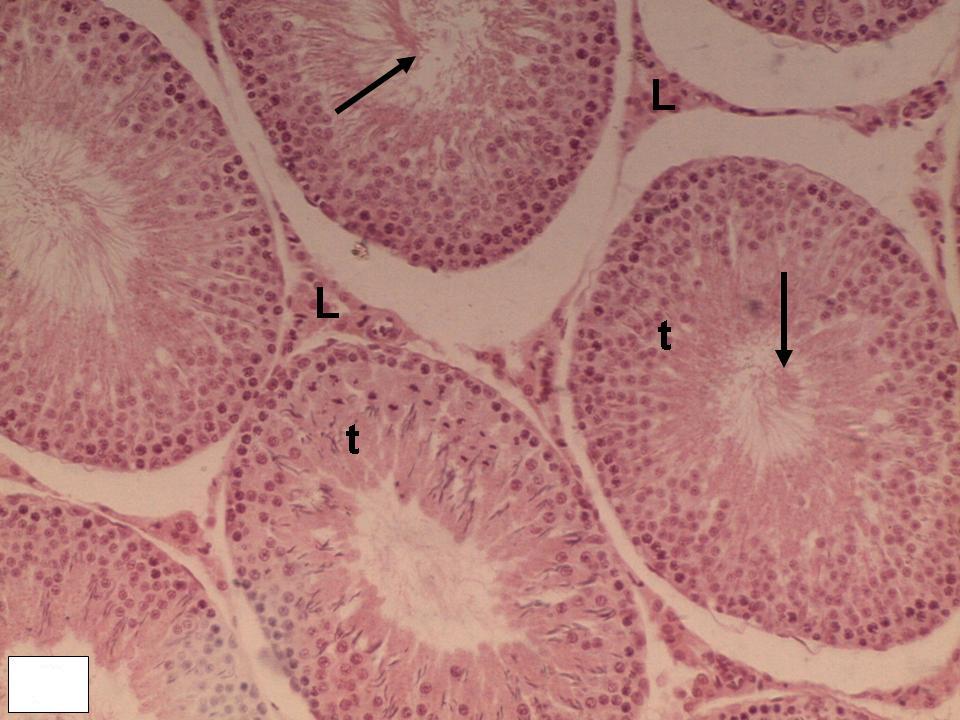
Degenerative changes were observed in both testes in the form of irregular
seminiferous tubules and degeneration of the germinal epithelium. A significant
decline in the number of PCNA-positive cells and in the sperm count of the exposed
group and a significant increase in the recovery group were observed. Moreover,
apoptosis was investigated in the present study, and a significant increase in the
expression of P53 was found, whereas it was significantly decreased in the recovery
group.
Conclusions
Mobile phone usage can decrease proliferation and increase apoptosis in
spermatogenic cells as well as decrease the sperm count. These changes are
reversible after stoppage of exposure to mobile phone radiation


