

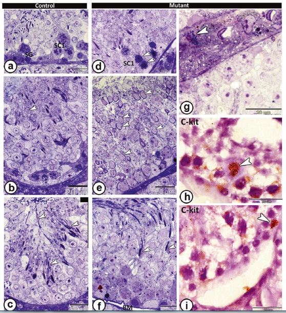
The role of retinoid-related orphan receptor, one of the transcription factors reported in testis, in testicular function is unclear, so this study was performed to evaluate the qualitative and quantitative changes in the testicular structure of RORα-deficient mice using light-, electron-microscopy, and immunohistochemistry. Among the most striking alterations observed in the testis of the mutant mice were hypospermatogenesis, marked reduction in volume proportions of interstitial tissues and number of Leydig cells, significant decrease in the diameter of seminiferous tubules and height of their epithelium, vacuolation in the epithelium of the seminiferous tubules with occurrence of mast cells, appearance of delay spermiation signs, and changes in sperm morphology. Moreover, the testis of mutant mice showed symplasts, in addition to appearance of multinucleated giant bromophenol-positive cells. ATPase activity was limited to spermatogonia and some primary spermatocytes, with higher alkaline phosphatase expression. Stronger vimentin reaction was immunolocalized to spermatogonia, spermatids, Leydig cells, and Sertoli cells. The expression of CD117 (C-kit, stem cell growth factor receptor) was limited to spermatogonia, primary spermatocytes, and Leydig cells. Seminiferous tubules showed overexpression of vascular endothelial growth factor (VEGF). Transmission electron microscopy examination of the mutant mice revealed abnormal Sertoli cells, hypertrophied spermatogonia, spermatocytes with degenerated mitochondria, and incompletely developed sperms. In conclusion, RORα is one of the essential proteins that regulate testicular structure.