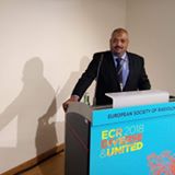
المعلومات الشخصية: medhat_ahmed

| الاسم بالكامل |
medhat_ahmed |
| البريد الإلكتروني |
medhat_ahmed@med.sohag.edu.eg |
| النوع |
ذكر
|
| الكلية |
كلية الطب |
| الدرجة الوظيفية |
استاذ مساعد |
| العنوان |
قسم الأشعه- كليه الطب |
| المنصب الحالي |
استاذ مساعد |
البيانات الأكاديمية
| التخصص العام |
الأشعة |
| التخصص الدقيق |
الأشعة التداخلية |
| عنوان رسالة الماجستير باللغة العربية |
دور الموجات فوق الصوتية والدولرالملون في تقييم قصور الشريان السباتى الفقارى |
| عنوان رسالة الماجستير باللغة الإنجليزية |
The role of color duplex ultrasonography in evaluation of vetrebrobasilar insufficiency. |
| عنوان رسالة الدكتوراه باللغة العربية |
تقييم الاجتثاث بالترددات الراديوية متعدد الأقطاب لعلاج سرطان الكبد في مرضى تليف الكبد |
| عنوان رسالة الدكتوراه باللغة الإنجليزية |
Assessment of multipolar radiofrequency ablation for the treatment of hepatocellular carcinoma in patients with cirrhosis |
| الوظائف الإشرافية و الإدارية |
منسق الجودة بقسم الأشعة |
| رقم الهاتف |
|
| Google Scholar |
|
| Linked |
|
| Research Gate |
|
| EKP بنك المعرفة المصري |
|
نبذه مختصرة
- CV
-
- I-PERSONAL STATUS:
- - Name: Medhat Ibraheem Mohammad Ahmad.
- - Address: Egypt. Sohag .Faculty of Medicine. Radiology department
- - Marital Status: Married and has three kids.
- - Language: Native arabic, Perfect English , good germany and french
- - Present Position: Lecturer of Diagnostic Radiology, Faculty of Medicine , Sohag University, Egypt .
- - Fax : (002) 093-602963
- - Email: mandw20022002@hotmail.com
- II- QUALIFICATIONS:
- M.B.B.Ch.: Faculty of Medicine, Sohag University, Sohag, Egypt. September,
- M.Sc. Radiology, Faculty of Medicine,Sohag University, Sohag, Egypt, in April, 2002, General grade: Very good.
- Radiology, Faculty of Medicine,Sohag University, Sohag, Egypt, in June, 2010, General grade: Very good.
- III- PREVIOUS POSTS:
- 1- House Officer (Rotating internship):
- At Sohag Faculty of Medicine, Sohag University Hospitals, ( from March 1997, through February 1998).
- 2- House Resident (Rotating internship):
- At Radiology Department ,Sohag Faculty of Medicine, Sohag University Hospitals, ( from March 1999, through December 2000).
- 3- House Resident (Rotating Internship) :
- At Radiology department, Cairo university Hospiatls (Al Kaser Al Eini), Cairo university, (from January 2001, through March 2002).
- 4- Assistant lecturer of radiology
- At Sohag Faculty of Medicine, Sohag University Hospitals, (from April 2002, through October 2004).
- 5- Resident
- At diagnostic and interventional radiology department, Johann Wolfgang Goethe University, Frankfurt, Germany. ( from November 2004, through December 2005).
- 6- Resident
- At diagnostic and interventional radiology department, Jean Verdier hospital, Bondy, Paris, France. ( from December 2005, through November 2006).
- 7- Assistant lecturer of radiology
- At Sohag Faculty of Medicine, Sohag University Hospitals, (from December 2006, through May 2010).
- 8- lecturer of radiology
- At Sohag Faculty of Medicine, Sohag University Hospitals, (from June 2010 till now).
- IV- Experience in:
- Gastrointestinal radiology :
- Conduct, supervise and interpret professionally the following imaging techniques:
- plain radiography and fluoroscopic contrast studies
- ultrasound
- CT
- MRI
- Accurately localize and take biopsy of abdominal and pelvic masses including lymph node biopsy
- Practice of safe and effective interventional techniques including the drainage of:
- hepatobiliary collections
- intra-abdominal collections
- pelvic collections
- Recognize the uses of radionuclide radiology in diagnosis of:
- GI bleeding
- tumours and metastases
- Recognize The role and limitations of ultrasound, CT, MRI and radionuclide radiology in the following conditions:
- The staging of GI and hepatobiliary cancers
- The investigation of abdominal trauma
- Identification of oesophageal, gastric, small bowel, and large bowel abnormalities
- Identification and categorization of peritoneal and mesenteric pathology
- Head and Neck/ENT/Dental radiology
- Professionally conduct, supervise and interpret the following imaging techniques
- plain radiography
- sialography
- dacryocystography
- ultrasonography including Doppler studies
- CT including: the primary diagnosis of benign and malignant lesions, staging of head and neck tumors, detection of skull base and neck tumors.
- MRI including: the primary diagnosis of benign and malignant lesions, staging of head and neck tumors, detection of skull base lesions, demonstration of cranial nerve anatomy and pathology.
- upper GI contrast studies including: barium swallow, Video fluorography, radionuclide imaging
- Accurately localize and biopsy neck masses and lymph nodes using ultrasound or CT scan
- Use the ultrasound or CT as a guide for drainage of head and neck collections
- Cannulate and dilate salivary gland ducts
- Cannulate and dilate lacrimal ducts.
- Musculoskeletal radiology
- Conduct, supervise and interpret competently, the following imaging .
- Plain radiography including primary care examinations, trauma cases,rheumatological disorders, general and pediatric orthopedic disorders.
- Ultrasonography of the joints and soft tissues including Doppler studies.
- Computed tomography including the use of CT for the primary diagnosis of benign and malignant pathology, the staging of Bone tumors, the detection of direct extension and metastatic spread .
- The use of MRI for the primary diagnosis of benign and malignant musculoskeletal pathology, the staging of tumors including the detec-tion of their direct extension and metastatic spread
- MRI and CT for the demonstration of spinal anatomy and pathology, joint anatomy and pathology, the investigation of rheumatological disorders and the investigation of trauma and sports injuries
- Radionuclide imaging
- Fluoroscopic procedures including CT an MR arthrography
- Accurately localize and biopsy the following:
- Soft tissue masses
- Bone masses
-
- Neuroradiology
- Perform and report cerebral angiogram
- Observe CT perfusion techniques
- Perform and report myelograms
- Perform and report carotid ultrasound including doppler
- Observe and interpret transcranial pediatric ultrasound
- Observe interventional neuro radiological procedures
- Observe MR functional brain imaging techniques
- Obstetric and Gynecological radiology
- Conduct, supervise and interpret the following imaging techniques: Ultrasound studies of the abdomen and pelvis including: transvaginal techniques, Doppler studies, contrast studies of the genitourinary tract, fluoroscopic studies of the genitourinary tract, CT and MRI for gynecological disease
- Accurately localize and take biopsy from pelvic and abdominal masses
- Use different imaging modalities for Drainage of abdominal-pelvic abscesses/collections
- Oncological radiology
- Conduct, supervise and interpret imaging techniques for the accurate diagnosis and staging of common tumors and their metastatic spread using: plain radiography, ultrasonography, computed tomography, magnetic resonance, radionuclide imaging andfluoroscopic procedures.
- Accurately localize and take biopsy from soft tissue masses and bone using ultrasound, or CT bone.
- Recognize The role of plain radiography, MRI, CT and nuclear medicine in the evaluation of an equivocal diagnosis of malignant lesions.
- Identify The indications and use of functional imaging.
- Observe The use of tumor specific agents and their application to cancer management.
- Observe The use of PET and CT PCT in cancer diagnosis and management.
- Recognize The value of imaging in therapy planning
- Pediatric radiology
- Conduct, supervise and interpret all imaging techniques used in the investigation of pediatric diseases including
- Plain radiography
- Ultrasonography and Doppler studies
- Computed tomography
- Magnetic resonance
- GI studies including identification of GI bleeding site
- Fluoroscopic procedures including: routine genitourinary
- investigations, routine gastrointestinal investigations
- Management of intussusception
- Disorders of swallowing
- Biopsy procedures
- Insertion of percutaneous nephrostomies
- Per-cutaneous aspiration and drainage techniques
- Thoracic radiology
- Conduct, supervise and accurately interpret the following imaging techniques
- Plain radiography specially for thoracic trauma
- Ultrasonography including demonstration of thoracic wall lesions and pleural effusions
- Computed tomography including identification and categorization of focal and diffuse lung disease
- CT pulmonary angiography
- Computed tomography of pleural lesions, thoracic wall and mediastinal lesions
- Magnetic resonance imaging
- Radionuclide radiology including ventilation/perfusion lung scan
- localize and take biopsy of the following:
- Thoracic wall lesions
- Pleural lesions
- Pulmonary lesions
- Mediastinal lesions
- Uroradiology
- Conduct, supervise and accurately interpret of the following imaging techniques:
- Digital radiography including intravenous urography, retrograde
- and antegrade pyelography, and cystourethrography
- Ultrasonography including Doppler studies
- Computed tomography of the urinary tract and pelvis including:
- unenhanced CT for detection urinary calculi, the staging of
- renal and vesical tumors
- CT-urography
- CT-angiography
- Magnetic resonance imaging of the urinary tract and
- pelvic organs
- Urodynamic imaging
- Imaging of renal transplant
- Perform safely and effectively the following interventional techniques:
- Biopsy of renal and retroperitoneal masses
- Drainage of collections
- Percutaneous nephrostomy
- Observe Antegrade pyelography, Renal cyst aspiration and ablation, Nephrolithotomy, Suprapubic vesical access and Transrectal
- Prostate biopsy
- 4.Staging of pelvic malignancy
- 5.Identification of pelvic floor disorders
- 6.Identification of renal and adrenal abnormalities
- Perform percutaneous nephrostomy
- Vascular and vascular intervention radiology
- Perform under supervision the following procedures
- Diagnostic arteriography
- Percutaneous peripheral angioplasty
- Percutaneous central venous access
- Thrombolysis
- IVC filter insertion
- Embolization
- Vascular stent insertion
- Alternative arterial access (e.g. Axilla)
- GI vascular dilatations and stent applications
- Transjugular liver biopsy
- Cardiac radiology
- Conduct, supervise and interpret the following imaging techniques to a high professional standard:
- Plain radiography
- Computed tomography
- Magnetic resonance imaging
-
- Breast radiology
-
- Use ultrasound in diagnosing breast diseases
- Use of radionuclide imaging in diagnosing and staging breast diseases
- Use of magnetic resonance imaging in evaluation of breast diseases
- Perform the following procedures
- Cyst aspiration
- Ultrasound guided fine needle aspiration cytology, localization and core biopsy
- Stereo tactic fine needle aspiration cytology, localization and
- core biopsy
- Mammographic guided localization for biopsy and treatment
- Ultrasound guided localization for biopsy and treatment
- Mammographic surgical specimen localization
-
- Special experience in:
- Image guided tumor ablation including.
- Radiofrequency ablation of liver and renal tumors.
- Percutaneous instillation of alcoholic and acetic acid in hepatic tumors
- V- PUBLICATIONS:
- Transarterial chemoembolization in the treatment of hepatoblastoma in children. Vogl TJ, Scheller A, Jakob U, Zangos S, Ahmed M, Nabil M. Eur Radiol. 2006 Jun;16(6):1393-6. 2005 Jul 8.
- Large (>or=5.0-cm) HCCs: multipolar RF ablation with three internally cooled bipolar electrodes--initial experience in 26 patients. Seror O, N'Kontchou G, Ibraheem M, Ajavon Y, Barrucand C, Ganne N, Coderc E, Trinchet JC, Beaugrand M, Sellier N.Radiology. 2008 Jul;248(1):288-96. 2008 May 15.
- Traitement percutane des varicoceles chez l’enfant et l’adolescent : criteres decisionnels pour l’embolisation par coil en complement de la sclerotherapie percutanee.Sellier1M.Ibrahim1Y.Ajavon1N.Zentar1N.Gault1O.Seror1V.Kazadjian2A.Wakim3F.Fayad1. Journal de Radiologie Volume 87, Issue 10, October 2006, Page 1239
- The impact of large vessel proximity on effectiveness of radiofrequency ablation of hepatocellular carcinoma: a controlled study . Seror O, N'Kontchou G, Muhammad M, Barrucand C, Tin Tin Htar M, Assaban M, Haddar D, Trinchet JC, Beaugrand M, Sellier N. J Radiol. 2007 Sep;88(9 Pt 1):1157-64.
- Impact of thrombocytopenia on radiofrequency ablation therapy of hepatocellular carcinoma in patients with liver cirrhosis.Medhat Ibraheem. The Egyptian Journal of Radiology and Nuclear Medicine 06/2013; 44(2):159–165.
- Expandable radiofrequency electrodes for ablation of centrally placed hepatocellular carcinoma. Medhat Ibraheem. The Egyptian Journal of Radiology and Nuclear Medicine. Volume 49, Issue 1, March 2018, Pages 92-98.
- Therapeutic response and risks of radio-frequency in treatment of hepatocellular carcinoma in patients with portal hypertension. Medhat Ibraheem. The Egyptian Journal of Radiology and Nuclear Medicine.Volume 48, Issue 4, December 2017, Pages 799-805.
- Radiofrequency ablation with monopolar cluster versus bipolar multipolar electrodes for the ablation of ⩾5cm hepatocellular carcinoma. Medhat Ibraheem. The Egyptian Journal of Radiology and Nuclear Medicine. Volume 47, Issue 4, December 2016, Pages 1443-1449.
- Sonographically Guided Radiofrequency Ablation of Subcapsular Hepatocellular Carcinoma MOHAMMED ZAKI, M.D. and MEDHAT IBRAHEEM, M.D. Med. J. Cairo Univ., Vol. 83, No. 2, March: 215-223, 2015
- The role of color duplex ultrasonography in evaluation of vetrebrobasilar insufficiency. Master thesis.
- Assessment of multipolar radiofrequency ablation for the treatment of hepatocellular carcinoma in patients with cirrhosis. Doctoral thesis.
- VI- REFERENCES:
- Prof. Mohammad Tharwat Al hawaige, Head of diagnostic Radiology, Radiology department, South valley university.
- Prof. Khaled Fawzy Hasan, Radiology department South valley university
- Prof. Ali Abdel Rahmaan, Clinical Oncology and nuclear medicine departments, South Valley university.
- Prof . Thomas J Vogl , Director of diagnostic and interventional radiology department, Johann Wolfgang Goethe University, Germany.
- Prof. Olivier Seror : Consultant of interventional Radiology , Jean Verdier hospital, Bondy, Paris, France.
- Prof. Nicolas Sellier : Head of Radiology department , Jean Verdier hospital, Bondy, Paris, France.
- VII- Core attitudes and behaviors
-
- Appreciate the legal and ethical framework within which radiology and general healthcare provision operate.
- Keep abreast of developments in information management relevant to radiology departments.
- Communicate effectively with patients and colleagues.
- Keep abreast of current trends and recent advances in clinical radiology.
- Strive for best practice in patient record keeping and the transfer of clinical data and images
-
