| Introduction |
- Epidemiology
- incidence
- rare
- accounts for 1% of all dislocations
- demographics
- more common in young or middle-aged males
- Pathophysiology
- typically result from a high-energy mechanism
- 25% may be open

- lateral dislocations more likely to be open

- Associated conditions
- associated dislocations

- associated fractures (up to 44%)

- with medial dislocation
- dorsomedial talar head
- posterior process of talus
- navicular
- with lateral dislocation
- cuboid
- anterior calcaneus
- lateral process of talus
- fibula
- Prognosis
- post-traumatic arthritis is common
- poorer outcomes associated with
- high-energy mechanisms
- lateral dislocations
- result from higher energy mechanisms
- open dislocations
- high risk of infection due to
- lack of muscle coverage
- poor vascularity of soft tissues
- difficulty cleaning contaminated joints
- concomitant fractures involving the subtalar joint
|
| Anatomy |
- Articulation

- inferior surface articulates with posterior facet of calcaneus
- talar head articulates with
- navicular bone
- sustenaculum tali
- lateral process articulates with
- posterior facet of calcaneus
- lateral malleolus of fibula
- posterior process consist of medial and lateral tubercles separated by groove for FHL
- Muscles
- talus has no muscular or tendinous attachments
- Blood Supply
- posterior tibial artery
- via artery of tarsal canal (most important and main supply)
- supplies most of talar body
- via calcaneal braches
- anterior tibial artery
- perforating peroneal arteries via artery of tarsal sinus
- deltoid artery (located in deep segment of deltoid ligament)
- supplies body
- may be only remaining blood supply with a talar neck fracture
|
| Classification |
- Anatomic
- medial dislocation
- most common (65-80%)
- due to lateral malleolus acting as strong buttress, preventing lateral dislocation
- results from inversion force on plantarflexed foot
- sustentaculum tali acts as fulcrum for the neck of the talus to pivot around
- foot becomes locked in supination
- associated with posterior process of talus, dorsomedial talar head, and navicular fractures
- reduction blocked by peroneal tendons, EDB, talonavicular joint capsule
- lateral dislocation
- more likely to be open
- results from eversion force on plantarflexed foot
- anterior process of calcaneus acts as fulcrum for the anterolateral corner of the talus to pivot around
- foot becomes locked in pronation
- associated with lateral process of talus, anterior calcaneus, cuboid, and fibula fractures
- reduction blocked by PT tendon, FHL, FDL
- anterior dislocation
- posterior dislocation
- total dislocation (extruded talus)
- talus is completely dislocated from ankle and subtalar and talonavicular joints
- results from continuation of forces required for medial or lateral dislocation with disruption of talocrural ligaments and extrusion of talus from ankle joint
- usually open
|
| Presentation |
- Physical exam
- foot will be locked in supination with medial dislocation
- known as "acquired clubfoot"
- foot will be locked in pronation with lateral dislocation
- known as "acquired flatfoot"
|
| Imaging |
- Radiographs
- recommended views
- findings
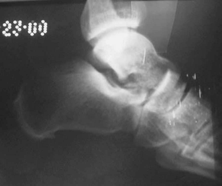
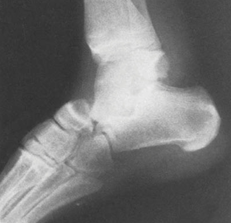
- medial dislocation
- talar head will be superior to navicular on lateral view
- lateral dislocation
- talar head will be collinear or inferior to navicular on lateral view
- CT scan
- indications
- perform following reduction
- findings
- look for associated injuries or subtalar debris
|
| Treatment |
- Nonoperative
- closed reduction and short leg non-weight bearing cast for 4-6 weeks
- indications
- 60-70% can be reduced by closed methods
- Operative
- open reduction
- indications
- open dislocations
- failure of closed reduction

- up to 32% require open reduction
- medial dislocation reduction blocked by lateral structures including
- peroneal tendons
- extensor digitorum brevis
- talonavicular joint capsule
- lateral dislocation reduction blocked by medial structures including
- posterior tibialis tendon is the most common
- flexor hallucis longus
- flexor digitorum longus
|
| Techniques |
- Closed reduction
- sedation
- requires adequate sedation
- reduction
- typical maneuvers include knee flexion and ankle plantarflexion
- followed by distraction and hindfoot inversion or eversion depending on direction of dislocation
- post-reduction
- perform a post-reduction CT to look for associated injuries
- Open reduction
- anesthesia
- approach
- dictated by direction of dislocation and associated fractures
- medial dislocation
- sinus tarsi approach to remove incarcerated lateral structures (EDB, etc.)
- lateral dislocation
- medial approach between tibialis anterior and posterior tibial tendon to remove medial structures (posterior tibialis tendon, etc.)
- may still require sinus tarsi/lateral approach to remove subtalar debris
- post-op care
- if joint stable
- place in short leg cast with non-weightbearing for 4-6 weeks
- if joint remains unstable
- place temporary transarticular pins or spanning external fixator
|
| Complications |
- Post-traumatic arthritis
- long-term follow up of these injuries show degenerative changes
- subtalar joint most commonly affected with up to 89% of patients demonstrating radiographic arthrosis (63% symptomatic)
- Stiffness
|