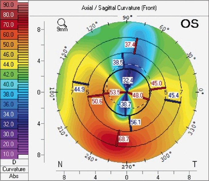
Pellucid marginal degeneration (PMD) is a rare ectatic corneal disease involving the inferior part of the cornea. It is difficult to differentiate between keratoconus (KCN) and PMD by slit lamp, especially in the detection of early and subclinical stages of the diseases. Corneal topography is the main diagnostic tool of PMD with characteristic diagnostic patterns “crab-claw” or “butterfly.” PMD could be mistaken as KCN, keratoglobus, and other peripheral thinning conditions such as Terrien marginal degeneration and Mooren's ulcer. Spectacles, soft and rigid gas permeable contact lens are the main visual correcting method in early stage of the disease. Different surgical techniques are available for PMD management; however, none of them were found to be effective, so further studies will be needed in the future.


