

This work was carried out to describe the morphological characteristics of the choanal slit of the turkey
through gross, light, and scanning electron microscopy. The choanal slit measures 27.62 mm long, and
constitutes 38.30 % of the total length of the palate. The edges of the narrow part of the choanal slit is
smooth rostrally but slightly thickened caudally due to the presence of 2-3 small papillae. The edge of
the wide part is thickened because of presence of 5-7 conical and wedge shaped papillae. SEM indicates
the presence of median fold within the choana, which represents the direct continuation of the median
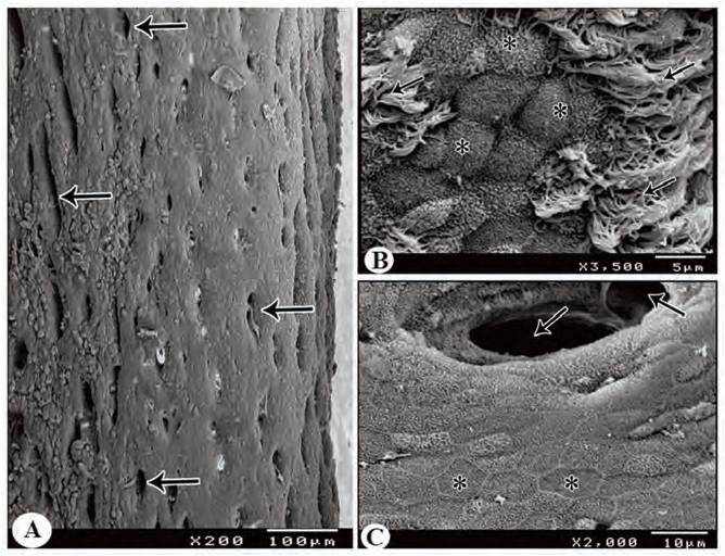
palatine ridge. After a short distance, this fold bifurcates into right and left folds. Several openings of
the palatine salivary glands are demonstrated on the palate at the level of the choanal slit. The epithelium
of the oral roof at the level of the choanal slit is stratified squamous epithelium showing intraepithelial
sensory corpuscles. This epithelium transforms at the edge of the choanal slit into
pseudostratified ciliated columnar epithelium that interrupted by intraepithelial mucous glands surrounded
by lymphatic infiltration and nodules. Altogether, this study provides inclusive information on
the macroscopic and microscopic morphological features of the choana in the turkey in comparing
with those of the other birds