
| Introduction |
|
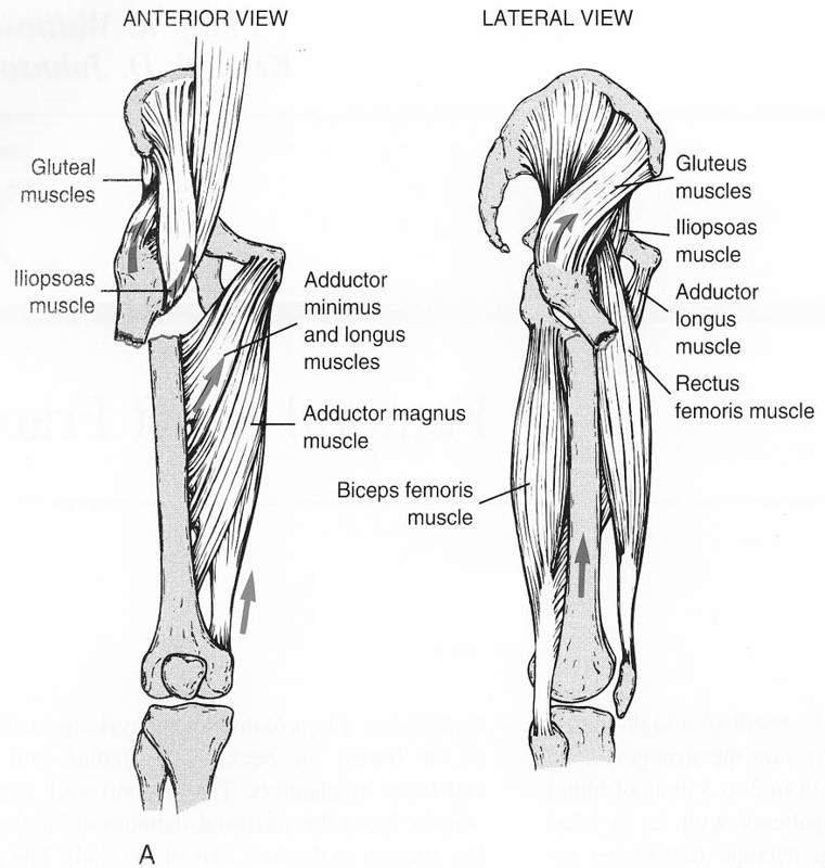
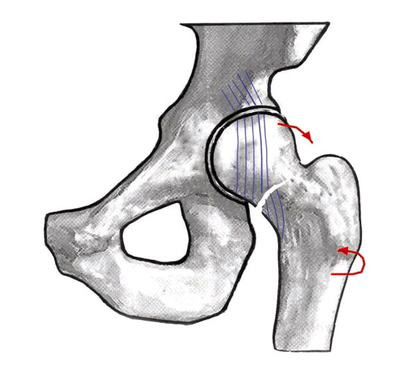
| Anatomy |
|


| Classification | ||||||||||||||||||
|
||||||||||||||||||
|
||||||||||||||||||
| Presentation | ||||||||||||||||||
|
||||||||||||||||||
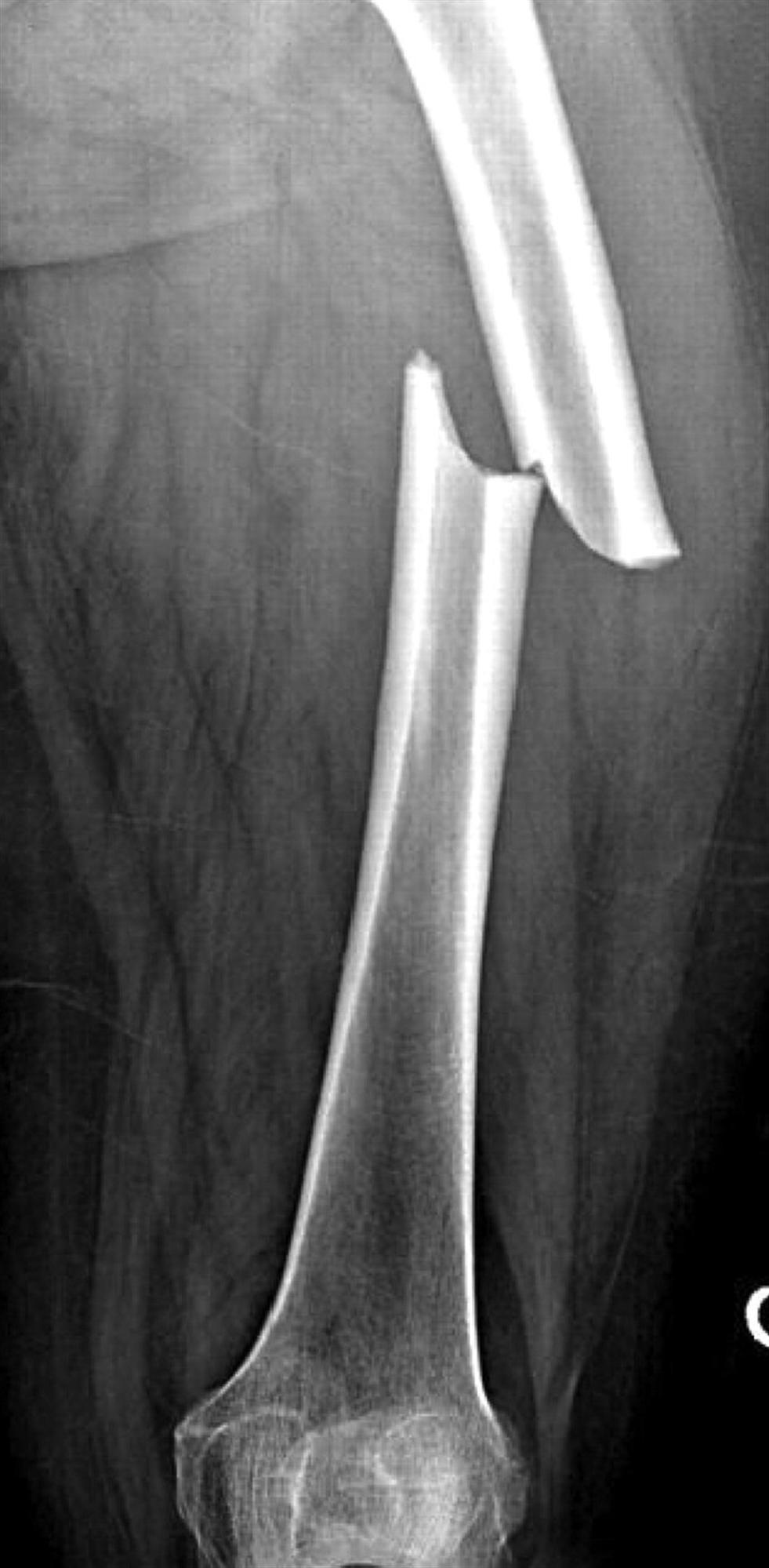
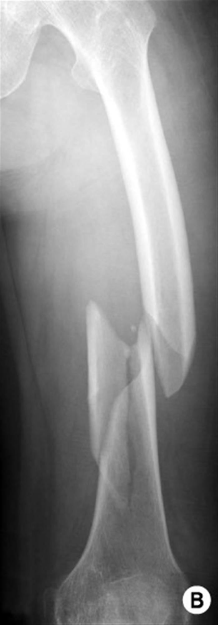
| Imaging | ||||||||||||||||||
|
||||||||||||||||||
| Treatment | ||||||||||||||||||
|
||||||||||||||||||
| Surgical Techniques | ||||||||||||||||||
|
||||||||||||||||||
| Complications | ||||||||||||||||||
|
||||||||||||||||||
| Introduction |
|
| Anatomy |
|
| Classification | ||||||||||||||||||
|
||||||||||||||||||
|
||||||||||||||||||
| Presentation | ||||||||||||||||||
|
||||||||||||||||||
| Imaging | ||||||||||||||||||
|
||||||||||||||||||
| Treatment | ||||||||||||||||||
|
||||||||||||||||||
| Surgical Techniques | ||||||||||||||||||
|
||||||||||||||||||
| Complications | ||||||||||||||||||
|
||||||||||||||||||
جميع البيانات و المستندات الموجوده لكل موقع من مواقع الاعضاء تقع صحتها قانونيا و أدبيا على العضو نفسه